 |
|
|
|
Swammerdam’s science
His life and work Nerve function Muscles Bees and ants "The Bible of Nature" Amazing drawings Techniques and microscopy Preformationism Swammerdam’s life Birth Death A fake “portrait” Science in society Empiricism and religion Mysticism and modern science Illustrations and their meaning Swammerdam in culture Swammerdam's world Friends and contemporaries Contemporary accounts On-line resources Under construction: Discussions of Swammerdam’s work A bibliography of Swammerdam's works Contact |
Over three centuries later, these drawings retain their power to amaze and give us an insight into the impact of the microscope not only on the reading public and the scientific world, but also on Swammerdam's vision of nature. The following selection of figures from the Bible of Nature gives some indication of his vision and his talent, and are all the more impressive given the fact that he had to invent the tools and techniques involved, and also did not know what to expect — none of these subjects had been studied before. A brief description follows each figure. 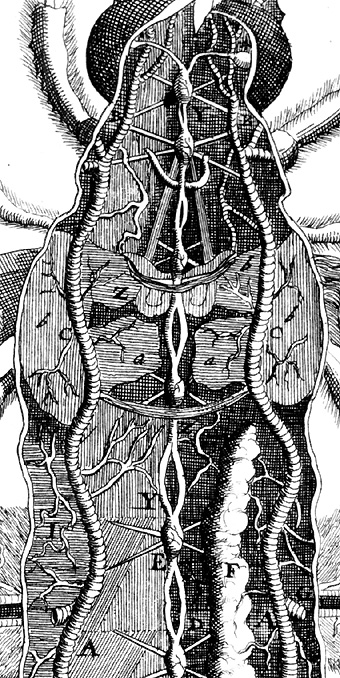 Dissection of a mayfly nymph showing nervous system (in the middle), trachea (the pipe-like structures on either sides) and muscles (hatched areas). Note the optic nerves branching out from the miniscule brain at the top. In the actual insect, the area shown here is about 8mm in length. 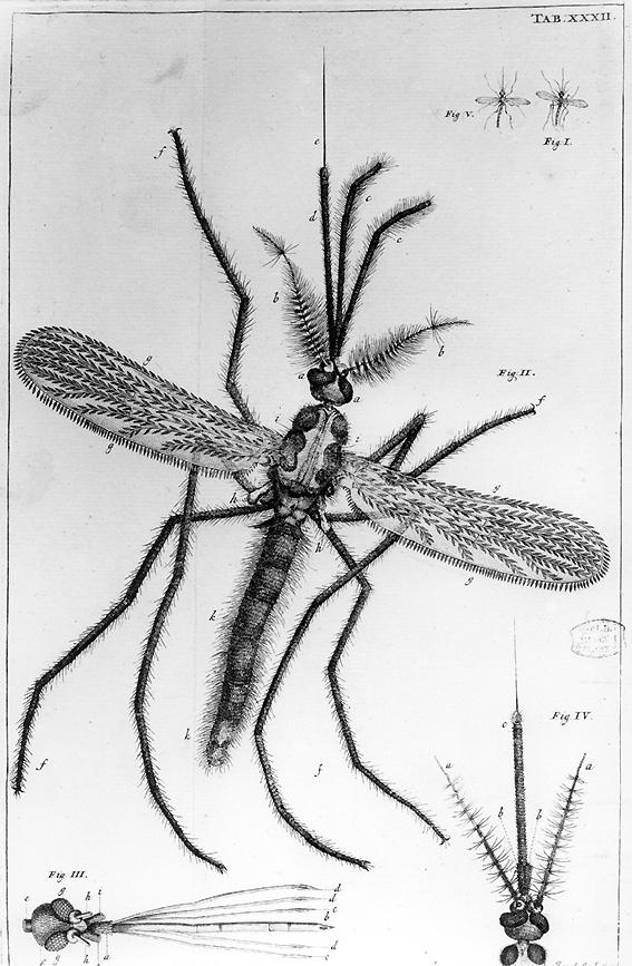 Drawing of a mosquito, adapted from the version that first appeared in Historia Insectorum Generalis (1669). This is a fine portrayal of the scales that cover the insect's wings (these were left out of the 1669 version) and of the complex stucture of the biting mouthparts. 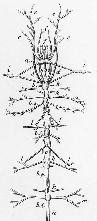 Dissection of the silkworm caterpillar nervous system. The small brain is marked "a". The nerves marked "e" are involved in smell and taste. 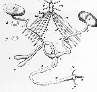 The bottom part of the same dissection. Swammerdam has included the testicles ("q") - these are present in the male caterpillar, even though he has no immediate use for them. Swammerdam noted the way the nerve "p" branches allows the vas deferens to pass through it at "s". Swammerdam wondered "whether this conduces to pleasure in this species of insect". The engraver has missed out a line on one of the nerves on the right, between "s" and "r". 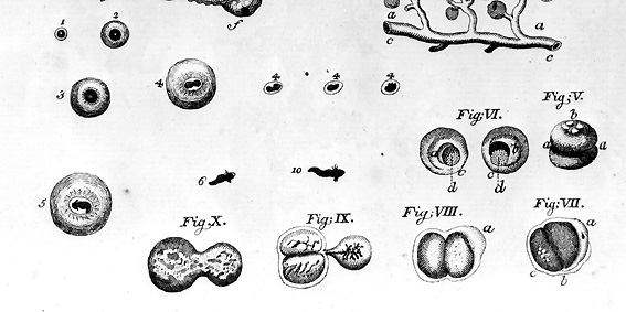 First cell divisions in a fertilised frog egg. These remarkable observations were the first to be made of thisivital initial stage of development. |